The Neck, What Bones And Cartilage Make It Up?
The neck is the part of the body that makes the transition between the trunk and the head. It is a vitally important structure, through which the blood vessels and nerves that are necessary for our survival pass.
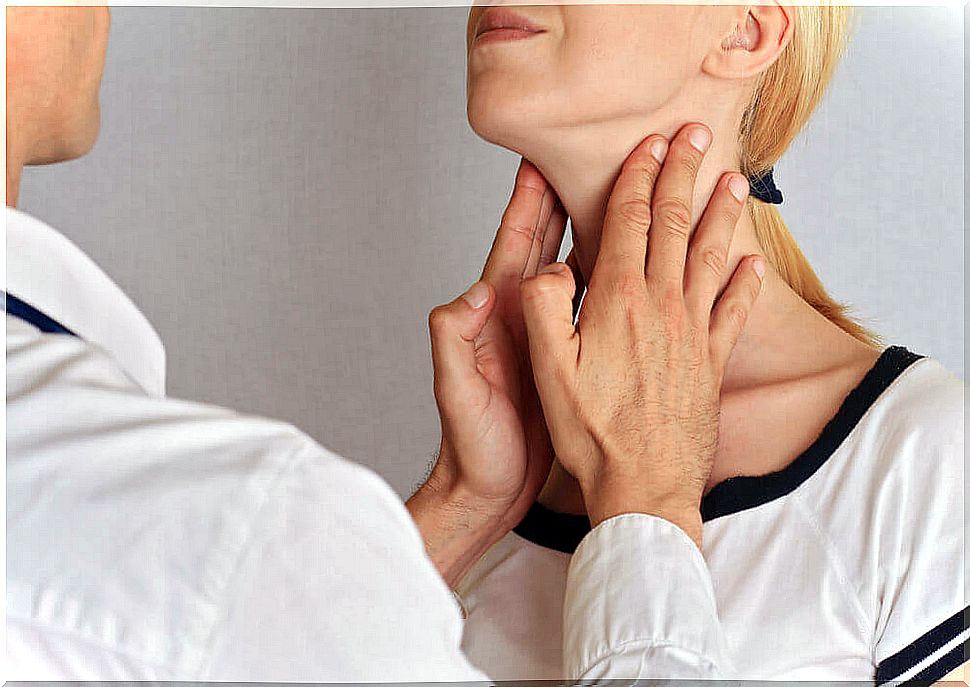
In the first instance, we could say that it is a thin and flexible area of the body that allows the mobility of the head. And although it does not seem like it, it is a vulnerable area, since any injury to it can damage the blood supply of the brain or its nerve transmissions.
The neck is a more complex structure than meets the eye, and in fact, it is one of the most complex in mammals, multiple studies reveal. It is made up of a series of bones, cartilage that articulates them, muscles, vessels and nerves.
Are you curious to know more? In that case, do not stop reading everything that we are going to tell you below.
How do we delimit the neck?

To facilitate the study of the neck, a series of superficial limits have been established that define it.
The neck begins at the lower edge of the jaw and the occipital bone, which is the base of the skull. From there, it extends to the clavicles and sternum in front. In the back, the neck reaches up to the C7 vertebra.
This structure contains many important elements that come together in a very small space. Among them, the following stand out:
- Pharynx.
- Larynx.
- Windpipe.
- Carotid arteries.
- Numerous nerves.
Next we will explain what is the framework that protects all these parts.
What are the bones of the neck?
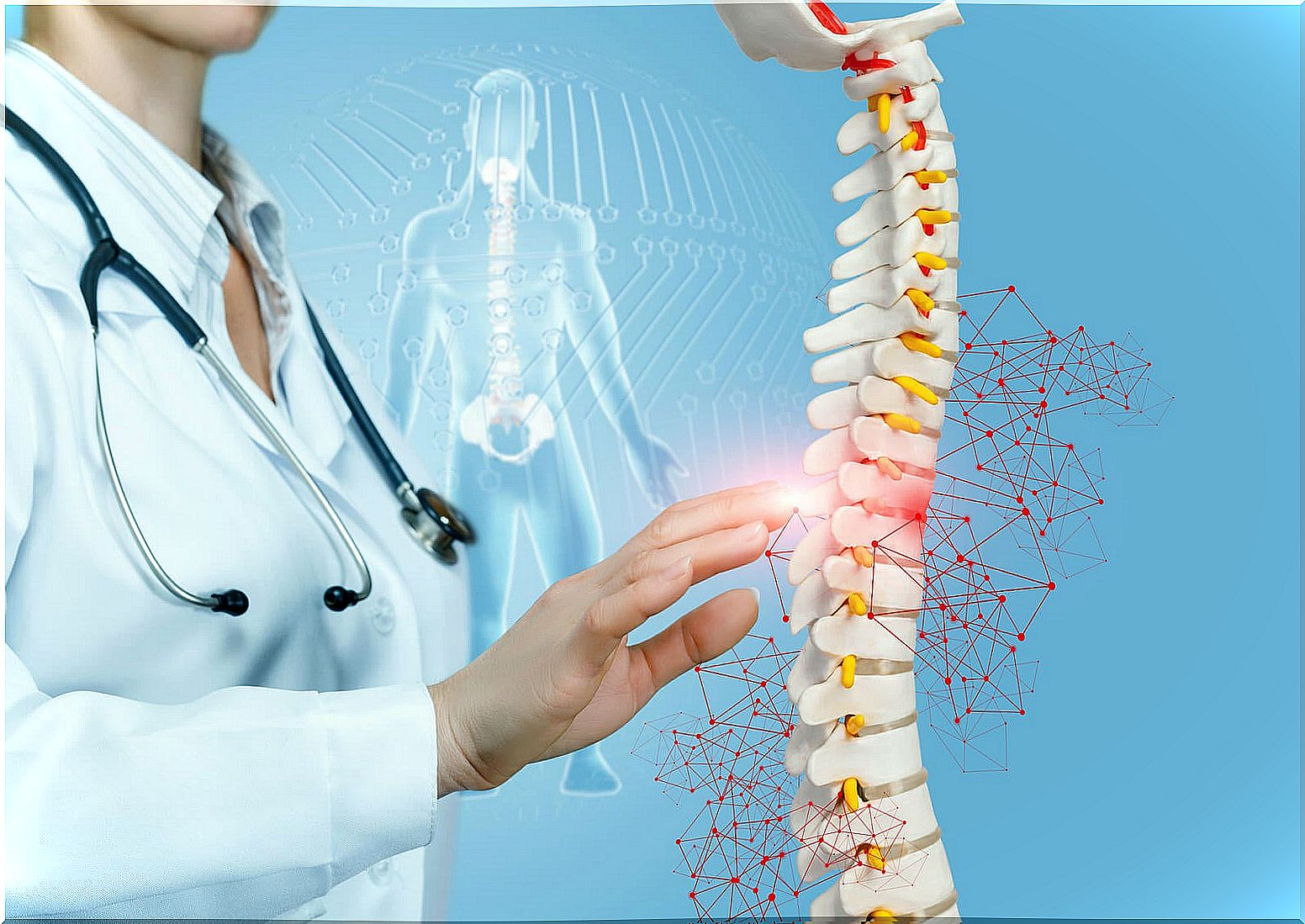
The skeleton of the neck is made up of the cervical vertebrae, the hyoid bone, the clavicles, and the sternum. The cervical part of the spine, that is, the neck, is made up of seven vertebrae.
In addition, there are intervertebral joints, which are what give it flexibility and movement. In fact, it is also a shock-sensitive structure that often suffers from pain.
Cervical vertebrae
The vertebrae are not all the same as each other. As we have mentioned, the neck is made up of seven vertebrae. From the third to the sixth they are all the same. They present a vertebral body and spinous process, which is the back part of the vertebra; they have a concave upper face and a convex lower face and are small in relation to the rest and a little flattened.
The first vertebra is called the atlas . It is a bone with the shape of a kidney ring, without a body or apophysis. It is formed by two lateral masses that are connected by arches, hence its ring shape. It is the vertebra that contacts the occipital bone.
The second vertebra or C2 is called the axis. What differentiates it from the rest of the cervical vertebrae is its process. It is the so-called odontoid process, which is a peg-shaped projection of your body.
Finally, the C7 vertebra differs from the rest also in its process. It is a spinous process , like those of the C3 to C6 vertebrae, but it is not bifid. This spinous process is longer than the other cervical ones and, because of this distinctive range, it is also called prominent.
Hyoid bone
The hyoid is a mobile bone that is located in the anterior part of the neck. It is at the level of the third vertebra, between the jaw and the thyroid cartilage. Interestingly, it does not articulate with any other bone. It is supported by a series of ligaments that run to the temporal bones of the skull, called stylohyoid ligaments. In addition, it is anchored to the thyroid cartilage.
What are the cartilages and ligaments of the neck?
First of all, we must mention the thyroid cartilage, the cricoid and the epiglottis. They form the front part of the neck, being part of the larynx and allowing breathing.
On the other hand, the cervical joints are formed by the discs that are interposed between the vertebrae and by a series of ligaments. The intervertebral discs have a central part called the nucleus pulposus and an external part called the fibrous nerve.
The anterior longitudinal ligament joins the vertebrae at the front. Similarly, there is a posterior longitudinal ligament. The yellow ligament connects the joints of two vertebrae followed by its posterior part.
The intertransverse and interspinous ligaments connect the vertebrae between their processes. Finally, we also find the supraspinatus ligament, which in its highest part forms the nuchal ligament.
As we have seen, the neck is a very complex part of the body that, to be understood, must be studied in depth. Its seven vertebrae, with their corresponding ligaments and intervertebral discs, are what allow it to be a flexible structure.