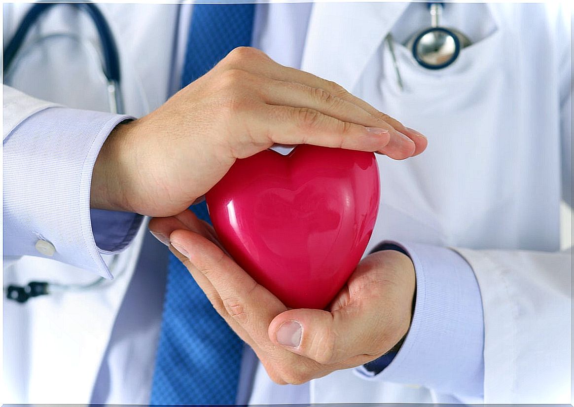
Nuclear Stress Test: What Is It Done For?
The nuclear stress test is one of the radiology studies used to evaluate the condition of the heart. Specifically, it allows you to see the blood flow of this organ. It is done at different times, both at rest and during exercise.
It can help diagnose problems with the coronary arteries, which are those that supply the heart. In addition, it serves to guide certain treatments. Therefore, in this article we explain what it consists of and everything you need to know about the nuclear stress test.
What is the nuclear stress test?
The nuclear stress test is a technique that has been developed recently. It allows you to observe how the blood flow of the heart is to check if there is any area that is not getting enough blood or is damaged.
This is done by measuring the flow at rest and during exercise. This is done by injecting a radioactive dye through a vein, ingested or even inhaled. This substance accumulates in the organs and emits gamma ray energy.
According to the RadiologyInfo specialists, images can be taken using special devices that allow us to observe the structure and function of the heart. The most widely used are single photon emission computed tomography (SPECT) and the gamma camera.
Multiple images are obtained. First during rest and later during exertion. This can be seen if the arteries of the heart are able to cope with the increased oxygen demand that occurs with exercise.
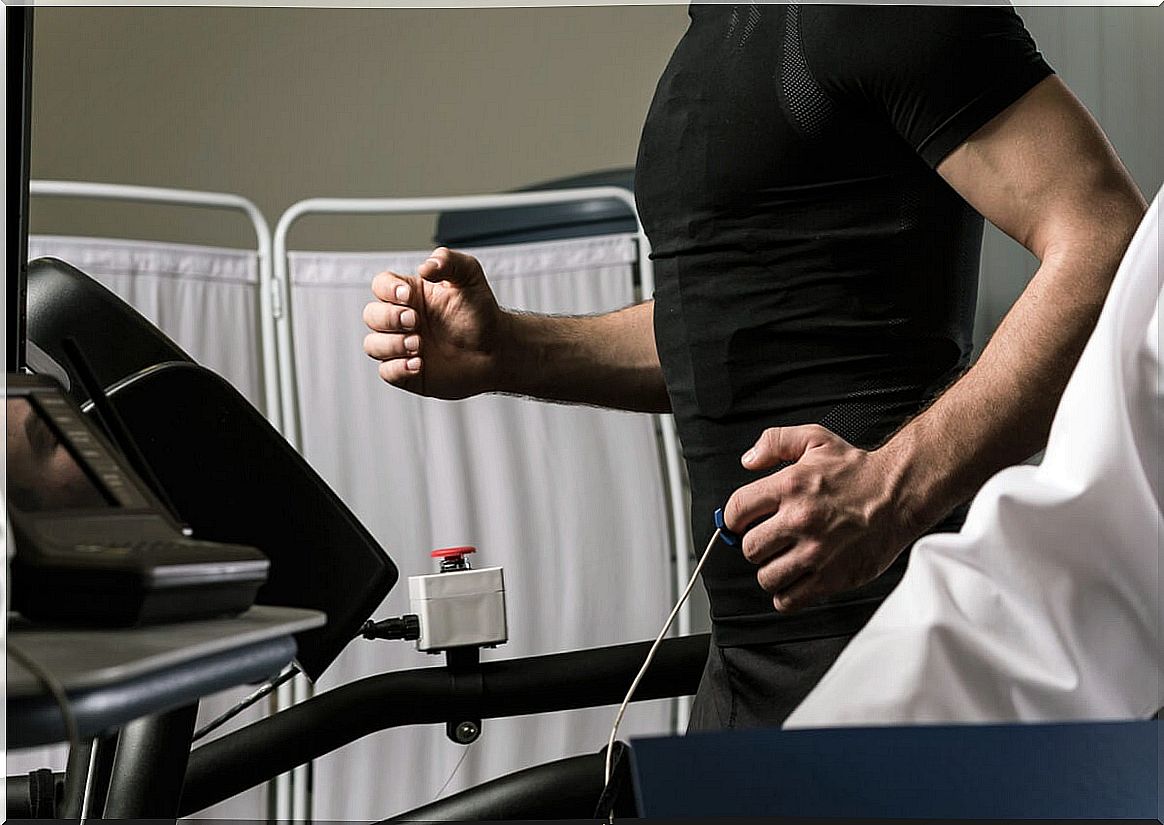
The nuclear stress test is performed in a controlled environment. Stationary bicycles or treadmills are usually used to generate this effort. It is a technique that, in addition to serving to diagnose, helps to better choose the treatment.
What is it done for?
According to specialists from the Mayo Clinic, one of the main purposes of the nuclear stress test is to diagnose coronary artery disease. They are the blood vessels that are responsible for carrying oxygenated blood to the heart.
These arteries are prone to damage as a result of atherosclerosis. This is a pathology that consists of the accumulation of cholesterol plaques and other substances on the arterial walls. These deposits prevent blood flow from passing normally.
When we exert ourselves, the heart needs to pump the blood faster and with more force to reach all the organs and tissues. In turn, the coronary arteries must also supply more blood to the heart for it to function properly.
Because of this, the arteries contract. If there is a plaque inside, it is possible that with vasoconstriction the blood flow is interrupted. This is evidenced by a nuclear stress test.
It can also be used to check whether, once the disease is diagnosed and treatment is in place, it is working effectively. The usual approach is usually based on drugs that help dilate the blood vessels, such as nitroglycerin.
Thus, the nuclear stress test is often recommended in people with angina pectoris. That is, when there is chest pain similar to that of a heart attack that appears in the face of efforts or stressful situations. It may also be indicated if there is an altered electrocardiogram.
How to prepare for the exam
Since what is desired is to observe the cardiac function, it may be necessary to avoid certain substances and medications before going. As Florida Cardiology specialists explain , it is important not to ingest caffeine or smoke before the test.
The doctor should be aware of any treatment that is being taken before. For example, certain inhalers used for respiratory conditions can alter the result. Therefore, it may be indicated to remove them before the nuclear stress test.
In the same way, as we have already pointed out, exercise is required during the procedure. Hence, the ideal is to go with sports and comfortable clothing. Electrodes are used that are glued to the skin, so no type of cream or lotion should be applied either.
How is the nuclear stress test performed?
The nuclear stress test usually takes hours. This is because you have to administer the radioactive drug and wait for it to be absorbed by the different tissues. Once 40 minutes have elapsed, images of the heart function at rest begin to be obtained. In order to get a complete idea, an electrocardiogram is also performed.
When the images have been taken at rest, the effort itself is carried out. Most often, an exercise bike or treadmill is used. Little by little the intensity of the exercise is increased and a point is reached where the heart rate is very high or symptoms begin to appear.
At this point, the radioactive drug is injected again. Images of the heart are taken again to see if any areas are not getting enough blood flow. As the dye is distributed with the blood, the least tinted areas will be those that have a circulation problem.
If at any time during the test the patient begins to feel very bad, it is stopped. It is common that, if there is a problem in the arteries, chest pain, dizziness or shortness of breath appear. After the nuclear stress test is completed, the images of rest and stress are compared.
Risks of the test
The nuclear stress test is a diagnostic method that usually does not have complications. However, that does not mean that it is risk-free. Derivative symptoms such as dizziness, nausea, anxiety or headache are common.
Similarly, it is usual for blood pressure to drop after exercise. Therefore, dizziness can be aggravated. Although it is more infrequent, in some cases arrhythmias appear. Some people may experience an allergic reaction to the radioactive drug.
Nuclear stress test results
To obtain the results of the nuclear stress test, the images of the heart obtained during rest and during exertion must be compared. This shows the areas that have not absorbed the radioactive dye well.
If you see an area that has not stained well during exercise, it is possible that there is a blocked coronary artery. In the same way, if the flow is also altered during rest, it could be that the arterial blockage is serious. The same is also true when there was a previous myocardial infarction.
The test of choice to check for damage is coronary angiography. It is a technique that allows us to observe the interior of the coronary arteries. If there is a blocked area, a stent can be placed to increase the caliber.
Nuclear stress test is useful and safe
This technique allows you to check the functioning of the heart and the state of its tissues. Not only does it help diagnose cardiac pathologies, but it can also serve to better guide treatment.
In addition, it is a safe test that has a low probability of complications. However, it is very important that the before and after instructions are followed in order to obtain adequate results.